Application Note – Bioaerosol N.8 – Legionella protocol according CDC USA

Introduction
Legionnaires’ disease was first recognised as a result of an outbreak of acute pneumonia that occurred at the Convention of the American Legion in Philadelphia USA in 1976. The route of infection has been recognised as the inhalation of small aerosol containing bacteria of the Legionella sp into the lungs of the host animal. The bacterium primarily responsible for this disease is Legionella pneumophila serogroup 1 Pontiac. It can result in typical Legionnaires’ disease an acute pneumonia with low attack rate and relatively high fatality rate or Pontiac fever, a mild non-pneumonia infection with a high attack rate.
Epidemiological studies have shown outbreaks to be mainly associated with cooling towers and hot water systems but also with whirlpool baths, clinical humidifiers in respiratory equipment, supermarket vegetable sprays, natural spa baths, fountains and potting composts.
Middle aged males, smokers and immuno-compromised patients are the most likely to succumb to this disease due to their reduced immune defence systems. The female population and children have a very low rate of incidence.
Monitoring of Legionella is often carried out as part of a planned programme of maintenance of a water system or cooling tower.
Air Sampling
In certain instances, it is therefore beneficial to demonstrate the presence of Legionellae in aerosol droplets associated with suspected reservoirs of the bacterium. Although not essential for the identification of reservoirs of disease-causing strains of Legionella, air sampling can better define the role of certain devices in disease transmission.
Sampling for biological contaminants in air can be used to determine the presence of a particular organism in an air sample, the number of viable organisms, the number of particles bearing organisms and the size distribution of particles. Before, initiating a sampling protocol, an investigator should determine which information will be collected, For each determination, the investigator should consider the type of sampler to be used, the probable concentration of organisms, sampling time, and environmental factors affecting the aerosol.
When obtaining air samples for Legionella, a primary objective is establishing the presence of the bacteria in aerosol droplets. The following protocol is directed toward this purpose. Although some information regarding particle size and numbers of viable bacteria can be calculated from these procedures, this information should be considered approximate.
Air sampler
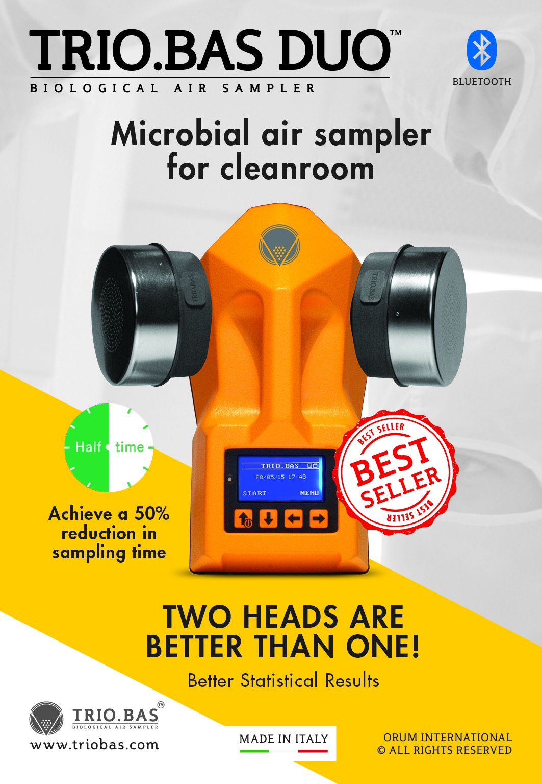
This Application Note deals with an impact on agar type portable air sampler.
Air is aspirated at a fixed speed for variable time through a cover which has been machined with a series of small holes of special design. The resulting laminar air flow Is directed onto the agar surface of a Contact Plate containing medium consistent with the microbiological examination to be made.
When the preset sampling cycle is completed, the plates are removed and incubated. The organisms are then visible to the naked eye and can be counted for an assessment of the level of contamination.
Sampling Protocol
1. Air sampler preparation
Be sure that the battery of the instrument is charged.
The aspirating head of the sampler should be sterilised by dry heating (160°C for 2 hours) or steam heating (autoclave at 121°C for 20 minutes) or isopropylic alcohol.
2. Air sampler positioning
2.1. HVAC (Heating Ventilation Air Conditioning) exit air-ducts The head of the air sampler should be positioned at about 30 cm from the exit of the air duct.
2.2. Shower, tap water, faucet, etc
The head of the air sampler should be positioned at about 30 cm from the water source. If necessary, fix the sampler to the floor tripod.
A background check should be performed with the water source turned off.
3. Volume of air
It is suggested to aspirate 500 litres of air for each cycle. The final result should be reported as Colony Forming Units (CFU) per 1000 litres of air (1 cubic metre).
Microbiological Media
Buffered charcoal yeast extract (BCY E_) agar
A. Composition
Norit SG charcoal 2.0 g
Yeast extract 10.0 g
ACES buffer 10.0 g
Ferric pyrophosphate (soluble) 0.25 g
L-cysteine HCl . H2O 0.40 g
Agar, bacto (Oxoid) 17.0 g
Potassium alpha-ketoglutarate 1.0 g
Distilled water 1.000 ml
Antibiotic components for media
1. GPAV medium
Glycine 0.3%
Polymyxin B 100 units/rnl
Vancomycin 5 ug/ml
Anisonlycin 80 ug/ml
2. PAV medium – GPAV medium without glycine
ABCYEαmedium
Bovine serum albumin (fractionV) 10 9/l
Dehydrated BCYEαagar base is available from
several commercial sources.
B. Media Preparation
1. Add ACES buffer to 940 mI of distilled water and dissolve in a 50°C water bath.
2. Add enough 1,0 N KOH (about 40 ml) to the buffer solution to bring the pH up to 6.9 and mix. (Do not use NaOH because it has been found to be inhibitory to Legionella pneumophila.)
3. Into a second flask, add charcoal, yeast extract, alpha-ketoglutarate, and agar. Mix the dry powders.
4. Pour the buffer solution into the second flask containing the dry powders and mix.
5. Carefully beat to dissolve the agar, then sterilise by autoclaving at 121°C for 15 minutes.
6. Immediately place the medium in 50°C water bath.
7. For complete medium, add membrane filtered solution of L-cysteine to the medium and mix thoroughly.
8. Add membrane-filtered soluble ferric pyrophosphate, and mix thoroughly. Do not mix iron and cysteine before adding to medium.
9. Adjust the pH of the medium to 6.9 at room temperature.
Since reagents may vary, each laboratory must determine the amount of KOH required. Hold the completed medium at 50°C, pour a 10- ml sample, and check the pH at room temperature. When necessary, adjust the completed medium with either 1.0 N K0H or 1.0 N HCl.
Note that the pKa of ACES buffer is 6.9 at 20°C and 6.8 at 25°C. Its pKa is affected by temperature (0,02 pH unit/°). However, once the agar has solidified, the pH does not appear to change with temperature but remains at 6.9.
10. For preparation of BCYEα + antibiotics, add membrane-filtered antibiotics and mix.
11. For BCYEα+albumin agar, dissolve the albumin in distilled water and filter sterilise before addition to the medium.
12. Dispense 20 mI per 15 X 100-mm Petri dish.
The medium must be mixed frequently during pouring to keep the charcoal particles suspended. After the medium has solidified, the plates should be stored in plastic bags in the refrigerator in the dark. They should be good for approximately 4 months, provided they pass quality control.
C. Precautions
1. The soluble ferric pyrophosphate must be kept dry and in the dark.
2. Fresh solutions of L-cysteine and ferric pyrophosphate, must be prepared each time. Since L-cysteine is a chelating agent, it must be added to the medium first. Both solutions must be added slowly. This is also true for adding 1,0 N KOH.
3. Do not use ferric pyrophosphate if its green colour becomes yellow or brown.
Quality Control of Media
Each batch of medium should be tested for pH and the ability to support the growth of L. pneumophila. Ideally, all media should be tested with a standardised tissue inoculum. However, since this material is not generally available, stock culture inoculum is recommended.
1. The pH of the medium must be 6.9 +/- 0.05. Measure with a pH meter. To check solid media, use a surface electrode (if available), or emulsify the agar from one plate in distilled water, and measure the pH of the emulsion. The pH is critical. If given lots of ingredients do not conform to the acceptable pH range adjust the pH of the completed liquid medium by adding 1,0 N HCl or 1,0 N KOH.
2. Check for support of growth in the following manner:
a. Prepare a standard stock culture inoculum by suspending L. pneumophila (log phase) in sterile distilled water to a concentration of109 cells/ml.
b. Inoculate the medium with 0,05 mI of the standard inoculurn. Streak the plates to obtain isolated colonies, and incubate at 35°C in air plus 2.5% C02.
c. Examine the media daily. On agar Petri dishes, growth should be present in the heavily inoculated area after 1 day. Isolated colonies should be macroscopically visible in 2 to 3 days. If cells in standard inoculum have been refrigerated or frozen, growth will be slower.
3. lf possible, selective media should be checked using an environmental water specimen containing a known concentration of legionellae and other bacteria.
Examination of Cultures for Legionellae
1. Examine all cultures after 72 to 96 hours incubation. Macroscopic examination should be done with a dissecting microscope andoblique lighting to detect bacterial colonies resembling Legionella. These colonies are convex and round with entire edges. The centre of the colony is usually bright white in colour with a textured appearance that has been described as “cut-glass like “ or speckled. The white centre of the colony is often bordered with blue, purple, green, or rod iridescence. Some species of legionellae produce colonies that exhibit blue-white or red autofluorescence. Further examination of primary isolation plates with long-wave UV light can easily detect these fluorescent colonies.
2. Aseptically pick each suspect colony onto
(a) BYCEα agar and
(b) BCYEα agar plate without L-cysteine [BCYEα (-)] or a BCYEα / BCYEα (-) biplate. Pick to – 4 representative colonies per sample.
3. Streak the inoculated portion of each plate with a sterile loop to provide areas of heavy growth, and incubate the plates for 24 hours.
4 . Bi-plates without growth are re-incubated an additional 24 hours. Colony picks that grow on BCYEα agar, but not BCYEα (-) agar, are presumptive Legionella species and must be examined further for definite identification using the direct fluorescent antibody (DFA) or slide agglutination test (SAT).
5. Since Legionella is a relatively slow-growing bacterium, negative plates should be re-incubated untiI 7days post-inoculation and then re-examined for Legionella colonies. Discard plates after the seventh day.
References
Center for Disease Control – Laboratory – Atlanta – USA.


